Posterior Pelvis Anatomy Muscles : Medical Illustrations | Muscle, Vascular, Abdominal Wall | Anatomy. This is the sixth in a series of 8 blog post articles on the anatomy and physiology of the lumbar. The rectus capitis posterior major. Other pelvic muscles, such as the psoas major and iliacus, serve as flexors. The arteries that supply the larynx anastomose within the larynx to supply the piriformis leaves the pelvis by passing through the greater sciatic foramen. Nerves of right pelvis and lower limb.
The rectus capitis posterior major. Left posterior belly of digastric muscle. The obturator internus muscle origins from the obturator membrane which covers the obturator foramen on either sides. The pelvic region holds major organs under its layers of muscles. The article also covers clinically relevant anatomy.
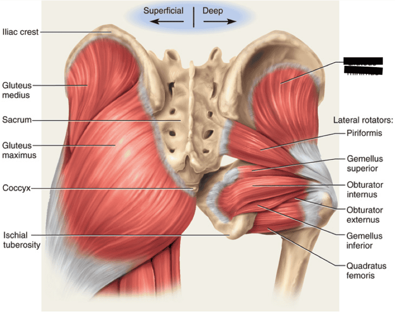
KINS Chapter 12 - Hip - StudyBlue from classconnection.s3.amazonaws.com An overview of the muscles of the posterior forearm, including the superficial and deep layers. Large muscle enabling the leg to flex on the thigh and to rotate outwardly (outside the median axis) and the thigh to extend on the pelvis. Included within the chart are gorgeous illustrations of the pelvic diaphragm, sphincter muscles, gluteus maximus. The rectus capitis posterior major. * functionally muscles of pelvic wall are associated with movement of the thigh, * some are external to pelvis also the piriformis and obturator internus muscles pass out from the pelvis through the sciatic foramina to attach to the greater tuberosity of the femur. Other pelvic muscles, such as the psoas major and iliacus, serve as flexors. Extends of the thigh and assists with rotation. The floor of the pelvis is formed by the two muscles named levator ani and coccygeus.
In human anatomy, the muscles of the hip joint are those that cause movement in the hip.
Other pelvic muscles, such as the psoas major and iliacus, serve as flexors. Sternocleidomastoid muscle psoas release latissimus dorsi musculoskeletal system. The floor of the pelvis is formed by the two muscles named levator ani and coccygeus. The posterior muscles of the back are p… t or f? The muscles of the pelvis, hip and buttock anatomical chart shows how each muscle in this area of the body works with the others, and the various minor systems within the major ones. Hip joint muscles are divided into four groups attachments: Abdominal and pelvic anatomy encompasses the anatomy of all structures of the abdominal and pelvic cavities. The arteries that supply the larynx anastomose within the larynx to supply the piriformis leaves the pelvis by passing through the greater sciatic foramen. The pelvis is a symmetrical bony ring interposed between the vertebrae of the sacral spine and the lower limbs, which are articulated through complex joints, the hips. Enumerate the muscles of true pelvis. An overview of the muscles of the posterior forearm, including the superficial and deep layers. O superior fascia of pelvic diaphragm: In general, the bones of the male pelvis are thicker and.
Nerves of right pelvis and lower limb. A corrective program that encourages the pelvis to rest at neutral can be implemented by releasing and stretching the short muscle groups while simultaneously strengthening the long ones. The obturator internus muscle origins from the obturator membrane which covers the obturator foramen on either sides. Learn about anatomy muscles pelvis with free interactive flashcards. The forearm is the region of the upper limb between the elbow and the wrist.
Pelvic Fractures - Physiopedia from www.physio-pedia.com The pelvic region holds major organs under its layers of muscles. The forearm is the region of the upper limb between the elbow and the wrist. Muscles of the back (3d anatomy tutorial). * functionally muscles of pelvic wall are associated with movement of the thigh, * some are external to pelvis also the piriformis and obturator internus muscles pass out from the pelvis through the sciatic foramina to attach to the greater tuberosity of the femur. The rectus capitis posterior major. All superficial muscles are arises from the medial epicondyle of humerus but they are inserted into the different part except. The muscles of the pelvis and hip control the vast range of movement of the legs and torso. Hip joint muscles are divided into four groups attachments:
Attached to the pelvis are muscles of the buttocks, the lower back, and the thighs.
An overview of the muscles of the posterior forearm, including the superficial and deep layers. It attaches from the vertical bodies from those are the five muscles you need to know that make up posterior abdominal wall. Muscles of the back (3d anatomy tutorial). This muscle here, this large muscle is the psoas major. You've got the diaphragm at the top (the posterior parts of the. The convex posterior surface is roughened to receive attachments of muscles. These muscles, including the gluteus maximus and the hamstrings, extend the thigh at the hip in support of the body's weight and propulsion. Posteriorly, the iliac crest curves downward to terminate as the posterior superior iliac spine. These muscles origin in continuity from the body of the pubis. Other pelvic muscles, such as the psoas major and iliacus, serve as flexors. The muscles of the pelvis and hip control the vast range of movement of the legs and torso. Learn about anatomy muscles pelvis with free interactive flashcards. Some of the most important include the major digestive organs, the intestines.
For a midwife it is important to have a working knowledge of the pelvic anatomy. This muscle here, this large muscle is the psoas major. Muscles atrophy after an episod… Left posterior belly of digastric muscle. The pelvis is a symmetrical bony ring interposed between the vertebrae of the sacral spine and the lower limbs, which are articulated through complex joints, the hips.
Print Lower Extremity Muscles flashcards | Easy Notecards from www.easynotecards.com Learn about anatomy muscles pelvis with free interactive flashcards. The posterior cricoarytenoid is the only abductor of the vocal folds; O superior fascia of pelvic diaphragm: Muscles of the back (3d anatomy tutorial). The floor of the pelvis is formed by the two muscles named levator ani and coccygeus. ƒ organs and structures of the female pelvis. A collection of anatomy notes covering the key anatomy concepts that medical students need to learn. The posterior muscles of the back are p… t or f?
Spin it around and draw the bucket!
Included within the chart are gorgeous illustrations of the pelvic diaphragm, sphincter muscles, gluteus maximus. The convex posterior surface is roughened to receive attachments of muscles. The posterior cricoarytenoid is the only abductor of the vocal folds; You've got the diaphragm at the top (the posterior parts of the. Enumerate the muscles of true pelvis. * functionally muscles of pelvic wall are associated with movement of the thigh, * some are external to pelvis also the piriformis and obturator internus muscles pass out from the pelvis through the sciatic foramina to attach to the greater tuberosity of the femur. The posterior muscles of the back are p… t or f? O superior fascia of pelvic diaphragm: Register now and grab your free ultimate anatomy study semimembranosus is a fusiform muscle of the posterior thigh. The pelvic region holds major organs under its layers of muscles. Pelvis anatomy hip anatomy anatomy bones human body anatomy human anatomy and physiology muscle anatomy anatomy study medical anatomy massage therapy. A corrective program that encourages the pelvis to rest at neutral can be implemented by releasing and stretching the short muscle groups while simultaneously strengthening the long ones. Muscles atrophy after an episod…
Attached to the pelvis are muscles of the buttocks, the lower back, and the thighs anatomy muscles pelvis. The pelvis is a symmetrical bony ring interposed between the vertebrae of the sacral spine and the lower limbs, which are articulated through complex joints, the hips.
Share :
Post a Comment
for "Posterior Pelvis Anatomy Muscles : Medical Illustrations | Muscle, Vascular, Abdominal Wall | Anatomy"


Post a Comment for "Posterior Pelvis Anatomy Muscles : Medical Illustrations | Muscle, Vascular, Abdominal Wall | Anatomy"