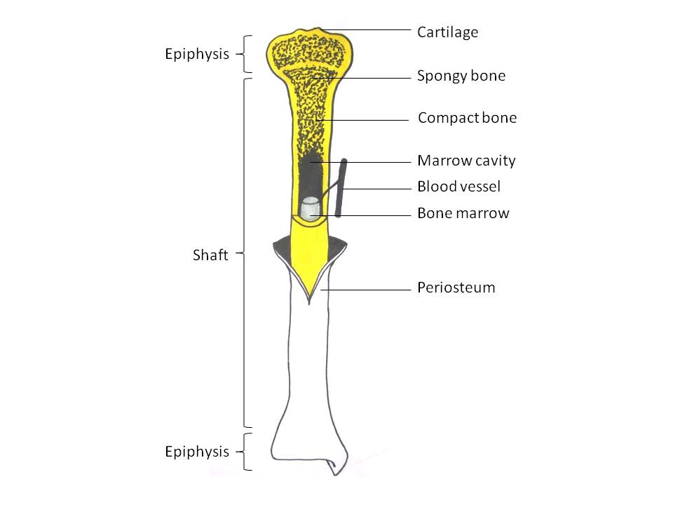
Long Bone Diagram Labled / E-Book 03 - Bone Structure: Compact Bone
Long Bone Diagram Labled / E-Book 03 - Bone Structure: Compact Bone. Human skeleton long bones of arms and legs. Each system contains haversian canals surrounded by concentric lamellae of bone tissue 48. Start learning with our skeleton diagrams, bone labeling exercises and skeletal system quizzes! Long bones are those that are longer than they are wide. Simple start to write diagrams.
Simple long bone diagram labeled : Study guide for students and teachers. Types of bones learn skeleton anatomy. Bone is found in the shafts of long bone and consists of various cylindrical units named as haversian system 47. They are one of five types of bones:

Long, short, flat, irregular and sesamoid.
.proximal epiphysis distal epiphysis medullary cavity compact bone articular cartilage spongy / cancellous bonel periosteum yellow bone marrow below, label the long bone to the right bonus: Human anatomy diagrams show internal organs, cells, systems, conditions, symptoms and sickness information and/or tips for healthy living. How to learn the human bones. Study guide for students and teachers. Each system contains haversian canals surrounded by concentric lamellae of bone tissue 48. Long, short, flat, irregular and sesamoid. Osteons of a long bone can be compared to a tree trunk. Short / long answer type questions. There are five types of human bones: When a human finishes growing these parts fuse together. Diaphysis proximal epiphysis distal epiphysis medullary cavity compact bone articular cartilage below, label the long bone to the right. The end of the long bone is the epiphysis and the shaft is the diaphysis. Diaphysis proximal epiphysis distal epiphysis medullary cavity compact bone articular cartilage.
Skull, clavicle, mandible, scapula, thorax, sternum, humerus, ulna, radius, carpus, phalanges (fingers), metacarpus, spine, pelvis, sacrum, femur, tibia, fibula, tarsus. Labeling portions of a long bone learn with flashcards, games and more — for free. Label the long bone purposegames. *free* shipping on qualifying offers. An easy and convenient way to make label is to generate some ideas first.
The medullary cavity contains red bone long bones follow the process of endochondral ossification where the diaphysis grows inside of cartilage from a primary ossification center until it.
The end of the long bone is the epiphysis and the shaft is the diaphysis. Simple start to write diagrams. Its lower end helps create the knee joint. The outside of the flat bone consists of a layer of connective tissue called the periosteum. This is an online quiz called long bone diagram labeling. Simple long bone diagram labeled : Hand | definition, anatomy, bones, diagram, & facts. Human skeletal diagram labeled bones college ruled composition notebook: Frontal skeleton orthopedic anatomy system publishing, castlecomer on amazon.com. Diaphysis proximal epiphysis distal epiphysis medullary cavity compact bone articular cartilage below, label the long bone to the right. Terms in this set (12). There are five types of human bones: And i dont mind which kind of bone cell it is, as long as its labeled.
The outer part of a long bone is made of compact bone. Human skeleton, the internal skeleton that serves as a framework for the body. The tough membrane covering the shaft of the bone. 12 photos of the long bone diagram labeled. *free* shipping on qualifying offers.

The medullary cavity contains red bone long bones follow the process of endochondral ossification where the diaphysis grows inside of cartilage from a primary ossification center until it.
Name the tissue which connects muscle to a bone. Skull, clavicle, mandible, scapula, thorax, sternum, humerus, ulna, radius, carpus, phalanges (fingers), metacarpus, spine, pelvis, sacrum, femur, tibia, fibula, tarsus. Long bone type in the upper arm. This framework consists of many individual bones and cartilages. Start learning with our skeleton diagrams, bone labeling exercises and skeletal system quizzes! Human skeletal diagram labeled bones college ruled composition notebook: Long bone structure diagram and definitions flashcards quizlet. Bone is found in the shafts of long bone and consists of various cylindrical units named as haversian system 47. Hand | definition, anatomy, bones, diagram, & facts. The outside of the flat bone consists of a layer of connective tissue called the periosteum. 12 photos of the long bone diagram labeled. Skeleton anatomy scheme with greater tubercle, deltoid. The outer part of a long bone is made of compact bone.
Post a Comment for "Long Bone Diagram Labled / E-Book 03 - Bone Structure: Compact Bone"